AI and Cancer Research ── Prostate ── 2026-10-18
Prostate cancer: treatment, trials and AI
- Abstract
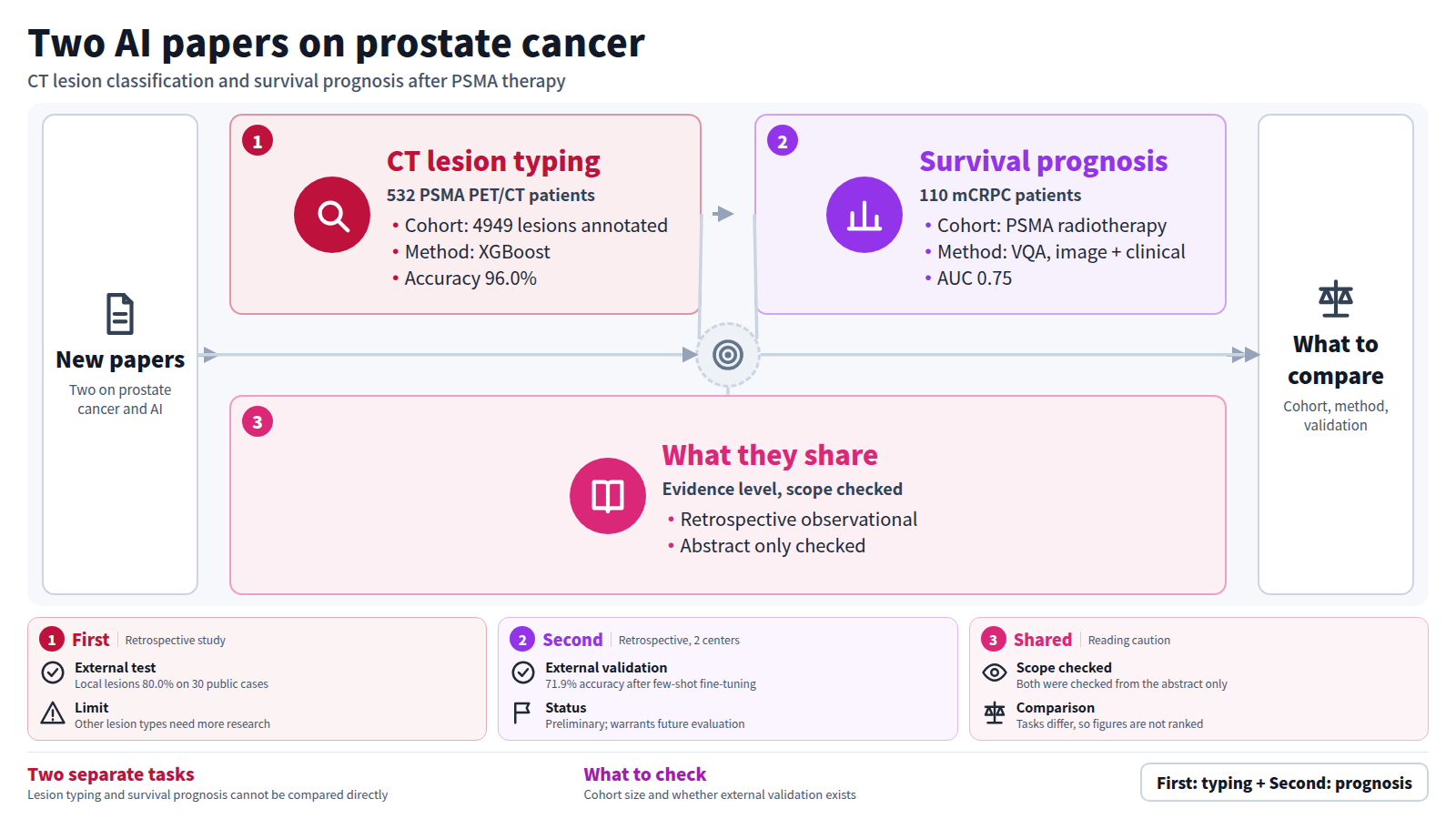
Accurate assessment of different types of prostate cancer (PCa) lesions is a crucial part of treatment planning. Following a computed tomography (CT) or a positron emission tomography (PET)/CT imaging scan, a significant amount of time is required from the clinicians to detect lesions and characterize them by their anatomical locations. To facilitate the development of new image analysis applications, we investigated the potential of automated classification of PCa lesions between bone metastases, lymph node metastases, local lesions, and other PCa lesions in CT images via machine learning (ML) and deep learning models. We annotated a total of 4949 PCa lesions by their type in a private dataset of 18 F-prostate-specific membrane antigen (PSMA) PET/CT images of 532 PCa patients and an external test set of publicly available PSMA PET/CT images from 30 patients. Using information of coordinates of the detected lesions, we automatically extracted 15 different features from the CT and the results of the automated 3D anatomical segmentation tool TotalSegmentator for each lesion, and trained several traditional ML models to classify lesions by their anatomical type. Additionally, we experimented with convolutional neural networks (CNNs) of different dimensionalities with CT and TotalSegmentator inputs. According to our results, the most accurate model was XGBoost with an overall accuracy of 96.0%. It identified bone metastases, lymph node metastases, and local lesions with sensitivities of 97.9±1.2%, 95.2±1.3%, and 93.1±2.8% in the five-fold cross-validation experiments of the primary data and 96.3%, 94.0%, and 80.0% on the external test set, respectively. Conversely, the CNNs underperformed the best ML models in distinguishing local lesions. To conclude, a simple ML model can accurately classify PCa lesions between bone, lymph node, and local lesions based on CT images, lesion locations, and automated anatomical segmentation data, but more research is required for distinguishing PCa lesions outside these categories.
Journal IF-equivalent: 3.9 (OpenAlex 2-year mean citedness, value as of 2026-10-13, retrieved 2026-10-15; not the official Clarivate IF)Reference: Rainio O, Vehmanen A, Li A, Jaakkola MK, Kemppainen J, Klén R. Investigation on Automatic Classification of Bone, Lymph Node, and Local Lesions in CT Images of Prostate Cancer Patients. J Digit Imaging Inform med. 2026 Oct 16 [Epub ahead of print]. doi:10.1007/s10278-026-02402-3.Checked: Abstract only - Abstract
Large language models show great promise for clinical decision support but generally lack medical imaging integration. Therefore, vision–language models have been proposed, but there is little evidence on their performance, particularly compared with conventional machine learning. Prostate-specific membrane antigen (PSMA)–targeted radiopharmaceutical therapy is a well-established treatment for metastatic castration-resistant prostate cancer (mCRPC), but it is still not known which patients will be good long-term responders. Therefore, this study proposes a physician-inspired visual question answering (VQA) framework that emulates the structured integration of PSMA-directed imaging and clinical data used in routine clinical assessment for overall survival prognostication.
Methods: As a proof of concept, we conducted a retrospective, multicenter study across 2 academic institutions that included 110 patients with mCRPC who were treated with either [177Lu]Lu-PSMA I&T or [177Lu]Lu-PSMA-617. Our VQA approach incorporates a 3-dimensional image encoder, the domain-specific language model RadBERT, and a cross-modal transformer to process baseline and interim (after 2 cycles) PET and SPECT images, clinical data, and clinical query. The model was trained to predict 24-mo survival, and performance was compared with state-of-the-art approaches. External validation was performed with the [177Lu]Lu-PSMA-617 cohort.
Results: The VQA model outperformed all state-of-the-art approaches while showing accuracy comparable to the query-free attention multimodal fusion model. Specifically, it achieved 81.8% accuracy (95% CI, 0.52–0.95) when integrating SPECT or PET images with prostate-specific antigen measurements. External validation of the VQA showed 71.9% accuracy (95% CI, 0.53–0.86) after few-shot fine-tuning, with further improvement over all state-of-the-art approaches in survival stratification. The VQA approach showed the closest calibration with the ground-truth survival distribution, with no significant differences observed (log-rank test, P = 0.76). Receiver operating characteristic analysis showed the highest area under the curve (0.75) among the evaluated models. Together, these findings suggest that the VQA approach demonstrated adaptability across different PSMA ligands while maintaining slightly improved yet comparable overall survival prognostic performance.
Conclusion: By adopting a physician-inspired assessment workflow, the VQA framework shows advantages in integrating imaging and laboratory data for clinical decision support compared with conventional machine learning. The preliminary results indicate that VQA-based approaches warrant future evaluation for survival prognostication in patients with mCRPC who are receiving PSMA radiopharmaceutical therapy.
Journal IF-equivalent: 6.9 (OpenAlex 2-year mean citedness, value as of 2026-10-17, retrieved 2026-10-18; not the official Clarivate IF)Reference: Kassar M, Hu J, Rauscher I, Gomes CV, Wendler T, Ziegler S, et al. Multimodal Visual Question Answering System for Survival Prognosis in PSMA-Targeted Radiopharmaceutical Therapy. J Nucl Med. 2026 Oct 15 [Epub ahead of print]. doi:10.2967/jnumed.125.271912.Checked: Abstract only