AI and Neurological Rare & Neuroimmune Diseases ── Multiple sclerosis ── 2026-10-16
Multiple sclerosis: treatment, trials and AI
- Abstract
Background: The use of generative artificial intelligence to create synthetic data is an emerging strategy to enhance real-world evidence.
Objective: To assess the validity of artificial intelligence-generated synthetic data in replicating data collected in the Italian Multiple Sclerosis and Related Disorders Register for the evaluation of progression independent of relapse activity risk under early intensive treatment and escalation treatment strategies.
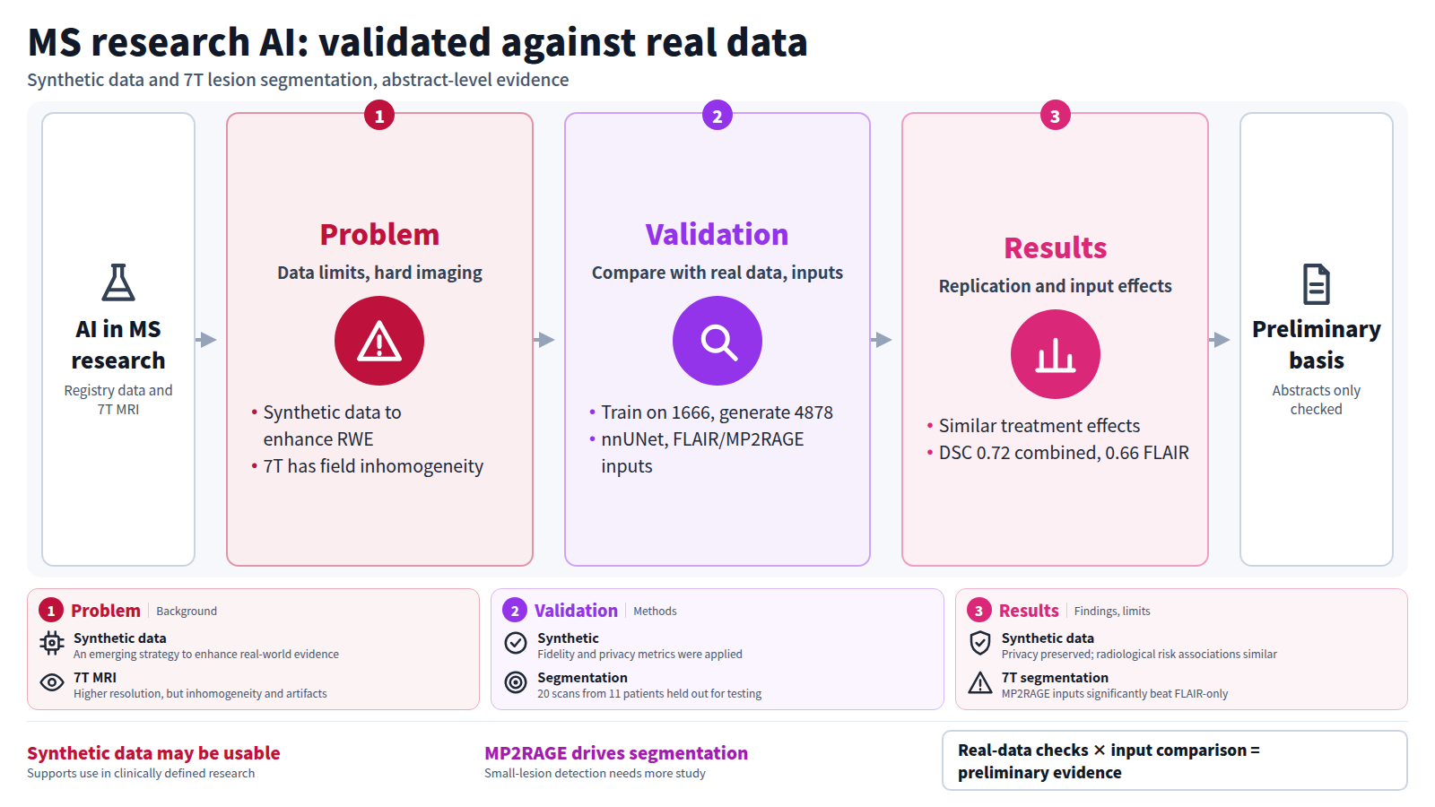
Methods: Artificial intelligence generative models were trained on a sub-cohort of 1666 Italian Multiple Sclerosis and Related Disorders Register patients to generate a synthetic dataset of 4878 patients. Synthetic vAlidation FramEwork, Clinical Synthetic Fidelity, and Nearest Neighbor Distance Ratio were applied. Comparisons between treatment strategies were conducted using both real and synthetic data, focusing on progression independent of relapse activity risk.
Results: The synthetic data demonstrated fidelity and privacy preservation. Treatment-effect estimates for escalation treatment versus early intensive treatment were similar across the real and synthetic datasets. Cox proportional hazards models showed that synthetic data preserved the robustness of the estimates for the risk of a first progression independent of relapse activity event. Associations with radiological risk factors were broadly similar across the real and synthetic datasets.
Conclusion: Artificial intelligence-generated synthetic data reliably replicated treatment-effect outcomes of Italian Multiple Sclerosis and Related Disorders Register data, supporting their potential use in clinically defined research settings.
Journal IF-equivalent: 3.5 (OpenAlex 2-year mean citedness, value as of 2026-10-15, retrieved 2026-10-16; not the official Clarivate IF)Reference: Iaffaldano P, D’Amico S, Lucisano G, Copetti M, Guerra T, Rocca MA, et al. Validation of generative artificial intelligence techniques for synthetic data generation in multiple sclerosis research: A comparison with real-world evidence from the Italian multiple sclerosis registry. Mult Scler. 2026 Oct 14 [Epub ahead of print]. doi:10.1177/13524585261492525.Checked: Abstract only - Abstract
Automated lesion segmentation can support rapid and reproducible quantitative assessment and long-term monitoring of multiple sclerosis. Ultra-high-field 7T MRI provides increased spatial resolution and lesion visibility but introduces challenges for automated analysis, including field inhomogeneity, susceptibility-related artifacts, and spatially varying image contrast. Although deep learning methods perform well at conventional field strengths, their application to 7T MRI remains underexplored. We evaluated nnUNet using FLAIR alone, MP2RAGE alone, and combined MP2RAGE+FLAIR. The dataset included 115 longitudinal 7T MRI scans from 57 patients with MS, with 20 scans from 11 independent patients reserved for testing. DeepMedic was included as a comparative architecture. On the test set, nnUNet achieved mean DSC values of 0.72, 0.71, and 0.66 for combined, MP2RAGE-only, and FLAIR-only inputs, respectively. No significant DSC differences were observed between nnUNet and DeepMedic. For both architectures, adding FLAIR to MP2RAGE did not show a statistically significant additional benefit in the present test cohort, whereas MP2RAGE-containing inputs significantly outperformed FLAIR-only input. The findings of the present study provide preliminary evidence suggesting that the observed voxel-level segmentation performance was primarily associated with the inclusion of MP2RAGE. However, lesion-level performance depended on lesion size, indicating further investigation of contrast-specific effects on small-lesion detection.
Journal IF-equivalent: 4.3 (OpenAlex 2-year mean citedness, value as of 2026-10-15, retrieved 2026-10-16; not the official Clarivate IF)Reference: Petrova A, Petrovic I, Berger M, Kravutske Y, Leinkauf J, Rumbak R, et al. Deep Learning-Based Multiple Sclerosis Lesion Segmentation at 7 Tesla: Comparison of Single- and Multi-Contrast MRI Inputs. J Imaging. 2026 Oct 14;12(10):510. doi:10.3390/jimaging12100510.Checked: Abstract only