AI and Cancer Research ── Brain ── 2026-10-09
Brain tumours: treatment, trials and AI
- Abstract
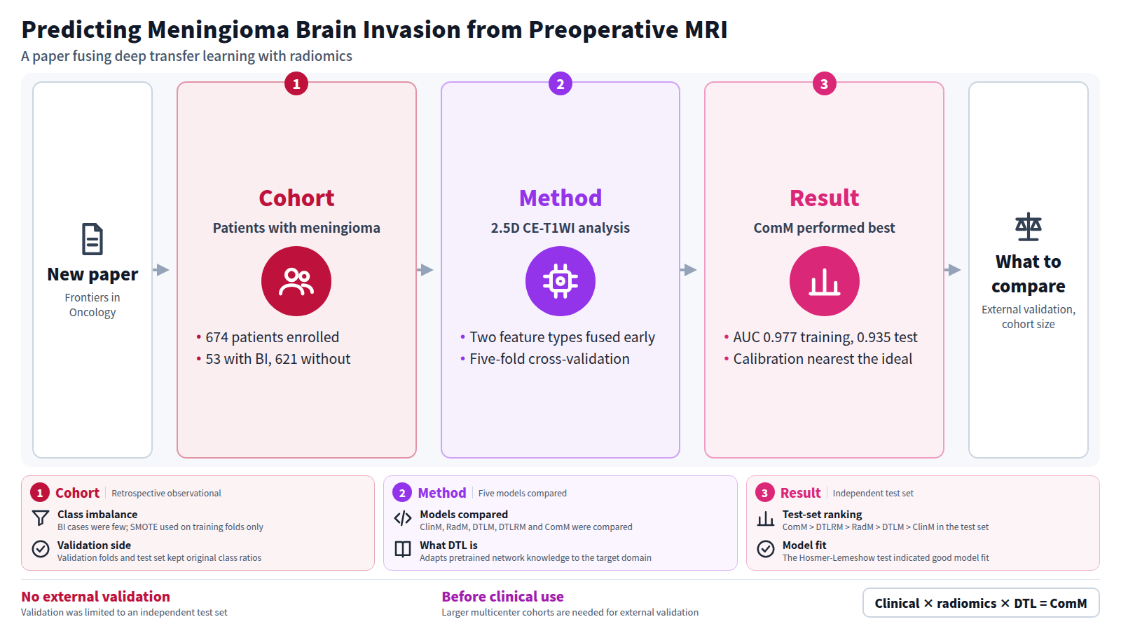
Background: Current preoperative assessment of meningioma brain invasion (BI) demands imaging analytical frameworks that optimize computational efficiency while capturing multidimensional tumor-pathology correlations. Building upon the inherent advantages of 2.5D MRI in synthesizing multiplanar biological data with manageable processing loads, this study innovatively integrates deep transfer learning (DTL), a technique bridging pretrained network knowledge with target domain adaptation, to systematically identify tumor-invasive biological signatures and develop an optimized predictive system using preoperative contrast-enhanced T1-weighted imaging (CE-T1WI).
Methods: This retrospective study enrolled 674 patients with pathologically confirmed meningiomas, including 53 with BI and 621 without BI. The cohort was stratified into a training set and an independent test set. Tumor regions of interest (ROIs) were delineated on CE-T1WI, and radiomics and DTL features were extracted. Radiomics and DTL features were fused via early fusion, followed by feature selection. Within the training cohort, five-fold cross-validation was performed, with SMOTE applied exclusively to the training portion of each fold; validation folds and the independent test set retained their original class distributions.
Results: The effectiveness of the models was compared using the area under the ROC curve (AUC). In the training set, ComM achieved an AUC of 0.977, while in the test set, its AUC reached 0.935. ComM demonstrated the best performance (training set: AUCComM > AUCDTLRM > AUCRadM > AUCClinM > AUCDTLM; test set: AUCComM > AUCDTLRM > AUCRadM > AUCDTLM > AUCClinM), showing excellent preoperative predictive capability for meningioma BI. The Hosmer-Lemeshow test indicated good model fit, and the calibration curve revealed that ComM was closest to the ideal curve.
Conclusion: ClinM, RadM, DTLM, DTLRM, and ComM based on 2.5D CE-T1WI all exhibited good performance in predicting meningioma BI. The clinical-radiomics-DTL fusion model (ComM) demonstrated the highest efficacy, suggesting that DTL provides additional features correlated with meningioma BI that differ from radiomics features. This model shows potential clinical utility for preoperative prediction; however, further external validation in larger multicenter cohorts is required before clinical implementation.
Journal IF-equivalent: 2.7 (OpenAlex 2-year mean citedness, value as of 2026-10-02, retrieved 2026-10-03; not the official Clarivate IF)Reference: Yuan D, Zhang J, Zhang C, Jing X, Feng Q, Han T. Non-invasive preoperative MRI-based deep transfer learning radiomics model for predicting meningioma brain invasion. Front Oncol. 2026 Oct 7;16:1935311. doi:10.3389/fonc.2026.1935311.Checked: Abstract only