AI and Neurological Rare & Neuroimmune Diseases ── Myasthenia gravis ── 2026-10-16
Myasthenia gravis: treatment, trials and AI
- Abstract
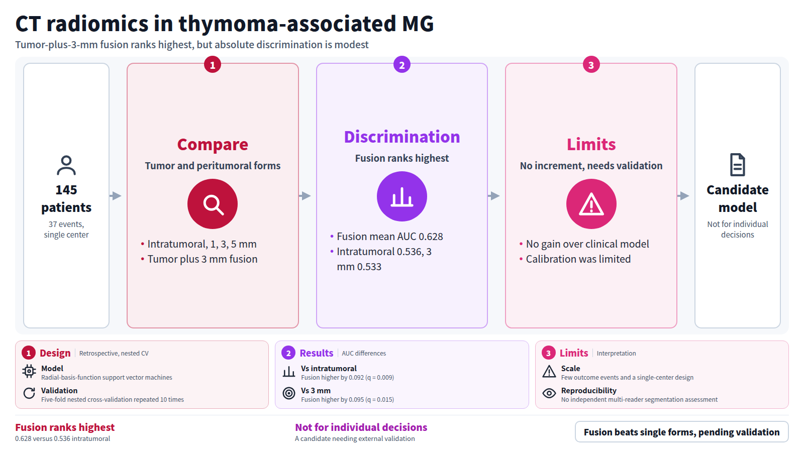
Background: Perioperative risk stratification in thymoma-associated myasthenia gravis remains challenging, and the optimal spatial extent for preoperative computed tomography (CT) radiomics is uncertain. We compared five three-dimensional tumor and peritumoral radiomic representations for discrimination of an in-hospital perioperative composite outcome.
Methods: This retrospective single-center study included 145 patients (37 events). Intratumoral, physical-distance peritumoral (1, 3, and 5 mm), and integrated tumor-plus-3-mm (VOIFusion3mm) representations were compared. Radiomic models used radial-basis-function support vector machines with five-fold nested cross-validation repeated 10 times. Prespecified paired comparisons used 2,000 patient-cluster bootstrap resamples with false-discovery-rate correction. Feature-exclusion, no-LASSO, boundary-perturbation, and clinical-increment analyses were also performed.
Results: The integrated tumor-plus-3-mm representation had the highest mean repeat-level out-of-fold area under the receiver operating characteristic curve (AUC) (0.628; 95% confidence interval (CI), 0.544–0.714), versus 0.536 for the intratumoral and 0.533 for the 3-mm peritumoral representation. AUC differences were 0.092 versus intratumoral (95% CI, 0.029–0.155; q = 0.009) and 0.095 versus 3-mm peritumoral (95% CI, 0.024–0.173; q = 0.015). VOIFusion3mm remained highest-ranked after feature exclusion and in the no-LASSO analysis. Mean AUC changed by − 0.093 after 1-mm inward erosion (95% CI, − 0.169 to − 0.015) and by − 0.004 after outward dilation (95% CI, − 0.028 to 0.019). Radiomics did not demonstrate incremental discrimination beyond the prespecified clinical model, and calibration was limited.
Conclusions: VOIFusion3mm showed higher internally evaluated rank discrimination than the intratumoral and isolated 3-mm peritumoral representations, but absolute discrimination was modest. Radiomics did not demonstrate incremental discrimination beyond the evaluated clinical models, and calibration did not support individualized risk estimation. VOIFusion3mm remains a candidate spatial representation requiring external validation and should not guide individual perioperative decisions. Interpretation is limited by the small number of outcome events, single-center design, and lack of independent multi-reader assessment of segmentation reproducibility.
Journal IF-equivalent: 3.8 (OpenAlex 2-year mean citedness, value as of 2026-10-15, retrieved 2026-10-16; not the official Clarivate IF)Reference: Chai Y, Lu C, Wang Z, Li Z, Li Z, Wang Y, et al. Integrated tumor–peritumoral CT radiomics for perioperative event discrimination in thymoma-associated myasthenia gravis: a retrospective cohort study. BMC Med Imaging. 2026 Oct 14 [Epub ahead of print]. doi:10.1186/s12880-026-02924-w.Checked: Abstract only