AI and Cancer Research ── Brain ── 2026-10-14
Brain tumours: treatment, trials and AI
- Abstract
Background: Accurate and timely classification is essential for brain tumor treatment planning. Classical machine learning (ML) with handcrafted features on moderate-sized datasets is under-represented, despite deep learning (DL) dominating medical image analysis.
Purpose: This study proposes a paradigm-agnostic comparison of classical ML and DL for brain tumor classification using a novel multi-domain feature framework, identifying which paradigm best balances accuracy, efficiency, and clinical use.
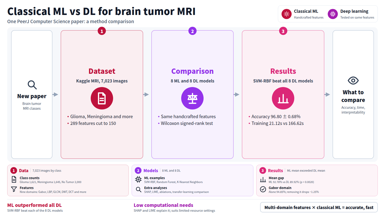
Methods: The Kaggle Brain Tumor magnetic resonance imaging (MRI) dataset (7,023 images; Glioma 1,621, Meningioma 1,645, No Tumor 2,000, Pituitary 1,757) was used. Nine feature domains—Gabor filters, local binary patterns (LBP), gray-level co-occurrence matrix (GLCM), discrete wavelet transform (DWT), discrete cosine transform (DCT), morphological, statistical, gradient, intensity gave 289 features, reduced to 150 by SelectKBest with the analysis of variance (ANOVA) F-test. Sixteen models were tested: 8 ML (Support Vector Machine with Radial Basis Function kernel (SVM-RBF), Random Forest, Gradient Boosting, Extra Trees, AdaBoost, K-Nearest Neighbors, Logistic Regression, Linear Discriminant Analysis) and 8 DL (Deep Multilayer Perceptron (Deep MLP), 1-Dimensional Convolutional Neural Network (1D-CNN), Bidirectional Long Short-Term Memory (Bi-LSTM), Gated Recurrent Unit (GRU), Convolutional Neural Network–Long Short-Term Memory (CNN-LSTM), ResNet-inspired, DenseNet-inspired, Autoencoder). The Wilcoxon signed-rank test assessed significance. Shapley Additive explanations (SHAP), Local Interpretable Model-agnostic Explanations (LIME), single-domain, progressive, leave-one-out (LOO) ablations, data-size sensitivity, and ImageNet transfer-learning comparisons were performed.
Results: SVM-RBF achieved 96.80 ± 0.68% accuracy, 96.73% precision, 96.63% recall, 96.65% F1, 99.63% Receiver Operating Characteristic Area Under the Curve (ROC-AUC), Cohen’s Kappa 0.957, Matthews Correlation Coefficient 0.957, outperforming all 8 DL models ( p ≤ 0.0039). DenseNet- and ResNet-inspired, trained on the same handcrafted features, reached 94.86 ± 0.76% and 93.95 ± 0.82%. The ML mean (92.78 ± 3.06%) exceeded the DL mean (89.92 ± 4.50%, p = 0.0020), with shorter training (21.12s vs . 166.62s, p = 0.0020). Per-class recall ranged 93.2–99.0%. Gabor was the most influential domain (single-domain 94.85%; SHAP 0.122; LOO drop −1.25%, p < 0.001).
Conclusions: Classical ML with multi-domain handcrafted features outperformed all 8 DL architectures with shorter training time, challenging DL’s assumed universality in medical imaging. The framework also raised DL accuracy by 1–16 percentage points over raw-image transfer learning, showing engineered features benefit both paradigms. With strong interpretability (SHAP, LIME) and low computational needs, the approach suits clinical deployment in resource-limited settings.
Journal IF-equivalent: 4.8 (OpenAlex 2-year mean citedness, value as of 2026-10-13, retrieved 2026-10-14; not the official Clarivate IF)Reference: Sethuraman R, Pandiarajan GS, Sadagopan P. Challenging deep learning universality: a complete evaluation of conventional machine learning and deep learning in classifying brain tumors using magnetic resonance imaging. PeerJ Computer Science. 2026 Oct 12;12:e4106. doi:10.7717/peerj-cs.4106.Checked: Abstract only