AI and Cancer Research ── Lung ── 2026-10-15
Lung cancer: treatment, trials and AI
- Abstract
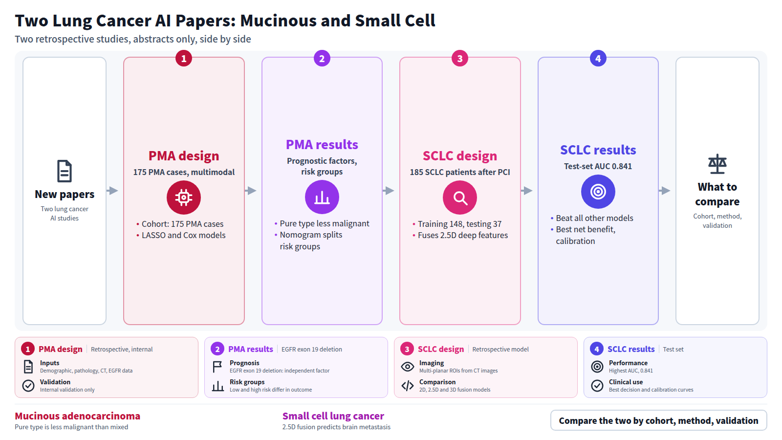
BACKGROUND: Pulmonary mucinous adenocarcinoma (PMA) represents a rare lung adenocarcinoma subtype, characterized by a lack of comprehensive pathological classification and prognostic factors. In this study, we propose a multimodal machine learning (ML) framework that integrates multiple data modalities to enhance PMA subtype classification and prognostic prediction. METHODS: This retrospective study enrolled 175 primary PMA cases with details of demographic, histopathological, computed tomography (CT) imaging, and epithelial growth factor receptor ( EGFR ) mutation data. Least absolute shrinkage and selection operator (LASSO) regularized logistic regression model was utilized for histological classification, and Cox proportional hazards model was employed for survival prediction with internal validation. RESULTS: Pure mucinous adenocarcinoma presented a higher prevalence of smaller radiographic or pathological tumor size, lower lobe localization, lower rates of lymph node metastasis and spread through air spaces (STAS), early pathological stage, carcinoembryonic antigen (CEA) ≤5 ng/mL, spiculated margin, Ki-67 low expression and lower frequency of EGFR exon 19 deletion (P=0.002, 0.003, EGFR exon 19 deletion as independent prognostic factors in PMA (P=0.04, 0.049, respectively). Moreover, Kaplan-Meier curves revealed significantly different outcomes between low- and high-risk groups stratified by the nomogram score (P<0.001). CONCLUSIONS: Pulmonary pure mucinous adenocarcinoma exhibited lower malignancy compared to the mixed mucinous and mucin-secreting type. The nomogram model developed and validated in this study exhibited promising efficacy in predicting histological subtype preoperatively and survival in patients with PMA, offering guidance for clinicians in diagnosis and prognosis prediction.
Journal IF-equivalent: 1.4 (OpenAlex 2-year mean citedness, value as of 2026-10-13, retrieved 2026-10-15; not the official Clarivate IF)Reference: Yang W, Chen C, Li L, Chen Z, Chen Z, Le H, et al. Integrating multimodal data for precise subtyping and prognostication in patients with pulmonary mucinous adenocarcinoma. Transl Cancer Res. 2026 Sep;15(9):708-8. doi:10.21037/tcr-2026-1241.Checked: Abstract only - Abstract
BACKGROUND: Brain metastasis remains an important clinical concern in patients with small cell lung cancer (SCLC), even after prophylactic cranial irradiation (PCI). Accurate identification of patients at high risk of brain metastasis after PCI may facilitate individualized surveillance and management. This study developed a fusion model based on two-and-a-half-dimensional (2.5D) multi-channel deep learning features, clinical features, and radiomics features to predict whether patients with SCLC will develop brain metastases following PCI. METHODS: This retrospective study included 185 patients with SCLC who underwent PCI and randomly divided them into a training set (N=148) and a testing set (N=37). A 2.5D multi-channel convolutional neural network (CNN) model was constructed based on multi-planar regions of interest (ROI) derived from CT images to extract 2.5D deep learning features. Based on the largest cross-section and three-dimensional (3D) volume of the tumor ROI, two-dimensional (2D) and 3D deep learning features were extracted, respectively. Subsequently, features from different dimensions were fused with clinical features and traditional omics features to construct the 2D_clinic_Rad, 2.5D_clinic_Rad, and 3D_clinic_Rad models, respectively. The performance differences between the 3D models and the clinical and traditional radiomics models were then compared. RESULTS: The 2.5D_clinic_Rad fusion model demonstrated the best performance in predicting brain metastases, achieving an area under the curve (AUC) of 0.841 on the test set, outperforming clinical models, traditional radiomics models, and 2D and 3D deep learning fusion models. Analysis of decision curves and calibration curves indicated that the 2.5D_clinic_Rad model demonstrated the highest clinical net benefit and the best calibration consistency compared to other models. CONCLUSIONS: The fusion model based on 2.5D_clinic_Rad can effectively predict the risk of brain metastasis in patients with SCLC following whole-brain prophylactic irradiation.
Journal IF-equivalent: 2.5 (OpenAlex 2-year mean citedness, value as of 2026-10-13, retrieved 2026-10-15; not the official Clarivate IF)Reference: Zhang X, Zhao L, Wang J, Zhang X, Liang X, Cao W, et al. Interpretable deep learning and radiomics for predicting the occurrence of brain metastases following prophylactic brain irradiation in small cell lung cancer. Transl Lung Cancer Res. 2026 Sep;15(9):281-1. doi:10.21037/tlcr-2026-0708.Checked: Abstract only