AI and Cancer Research ── Liver ── 2026-10-15
Liver cancer: treatment, trials and AI
- Abstract
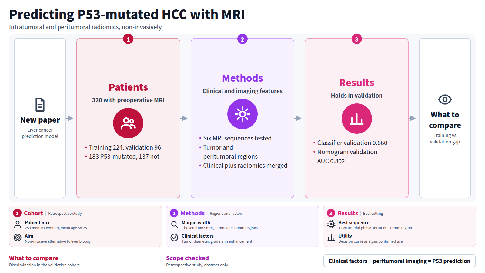
BACKGROUND: Hepatocellular carcinoma (HCC) is one of the leading causes of cancer-related mortality worldwide, and TP53 mutation acts as a trunk mutation driving tumor progression. However, TP53 mutation status can currently be determined only by invasive liver biopsy. This study applies multiregional radiomics features from magnetic resonance imaging (MRI) to predict P53-mutated HCC and offers a non-invasive tool for identifying this invasive subtype. METHODS: In this retrospective study, data from 320 patients who underwent preoperative MRI were analyzed and divided into a training cohort (TC; n=224) and a validation cohort (VC; n=96). Clinical risk factors were identified using stepwise regression to construct predictive models. We evaluated three machine learningclassifiers across six MRI sequences, and the least absolute shrinkage and selection operator method was applied to select the best IntraPeri fusion model from four regions (whole tumor, IntraPeri_9mm, IntraPeri_11mm, and IntraPeri_13mm) to establish a radiomics model. The predictive nomogram for P53-mutated HCC integrated these clinical predictors with discriminative radiomics features, and its performance was assessed using receiver operating characteristic analysis, calibration, and decision curves. RESULTS: Among 320 patients (259 men and 61 women; mean age, 58.25±11.51 years), 183 had P53-mutated HCC and 137 had non-P53-mutated HCC. The largest tumor diameter (LTD), Edmondson-Steiner grade, and rim arterial phase hyperenhancement were identified as independent predictors of P53-mutated HCC. The ExtraTrees classifier based on radiomics features from the optimal MRI sequence [T1-weighted imaging-arterial phase (T1WI-AP)] within the IntraPeri_11mm region achieved strong predictive accuracy [area under the curve (AUC) TC =0.805; AUC VC =0.660]. Predictive nomogram integrating clinical and radiomics features demonstrated excellent performance in both the TC [AUC =0.815; 95% confidence interval (CI): 0.758-0.872] and VC (AUC =0.802; 95% CI: 0.713-0.890). Decision curve analysis further confirmed its clinical utility. CONCLUSIONS: Nomogram combining clinical model with IntraPeri_11mm radiomics may serve as effective predictors of preoperative P53 mutation status in HCC.
Journal IF-equivalent: 1.4 (OpenAlex 2-year mean citedness, value as of 2026-10-13, retrieved 2026-10-15; not the official Clarivate IF)Reference: Weng J, Yao D, Zeng Q, He Y, Yang C, Yang B. Magnetic resonance imaging-based radiomics for predicting P53-mutated hepatocellular carcinoma: intratumoral and peritumoral analysis. Transl Cancer Res. 2026 Sep;15(9):709-9. doi:10.21037/tcr-2026-1579.Checked: Abstract only