AI and Cancer Research ── Kidney ── 2026-10-17
Kidney cancer: treatment, trials and AI
- Abstract
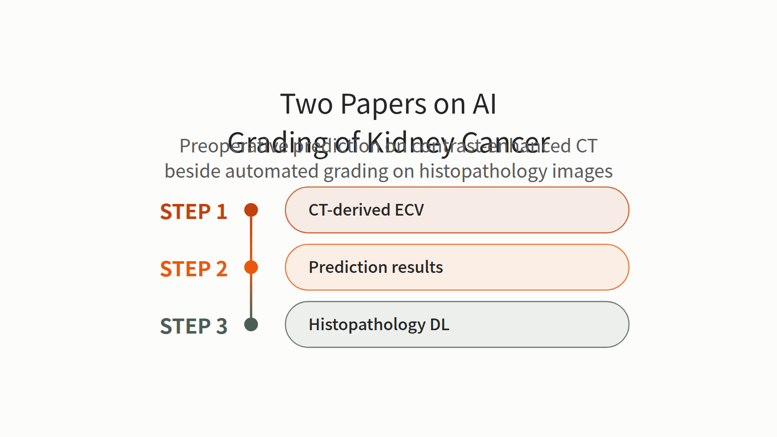
Objective: To develop and validate an interpretable machine learning (ML) model that integrates extracellular volume fraction (ECV) derived from contrast-enhanced CT with clinical and imaging features for preoperative prediction of World Health Organization/International Society of Urological Pathology (WHO/ISUP) grade in clear cell renal cell carcinoma (ccRCC). Materials and methods A retrospective cohort of 263 patients with pathologically confirmed ccRCC from two institutions was analyzed. Patients from Hospital A (n=184) were randomly divided into training (n=128) and internal validation (n=56) cohorts, while patients from Hospital B (n=79) served as an independent external validation cohort. CT-derived ECV, arterial enhancement fraction (AEF), and conventional imaging features were extracted. Six machine learning models (Logistic Regression [LR], eXtreme Gradient Boosting [XGBoost], Gaussian Naive Bayes [GNB], Support Vector Machine [SVM], K-Nearest Neighbors [KNN], Light Gradient Boosting Machine [LightGBM]) were constructed, and the best-performing model was identified based on bootstrap resampling. Model interpretability was assessed using SHapley Additive exPlanations (SHAP).
Results: Tumor size (low-grade: 4.99 ± 1.98 cm; high-grade: 6.20 ± 2.72 cm, P = 0.002), ECV (low-grade: 0.46 ± 0.09; high-grade: 0.57 ± 0.12, P<0.001), and AEF (low-grade: 1.37 ± 0.24; high-grade: 1.56 ± 0.25, P<0.001) were significantly higher in high-grade ccRCC, while hematocrit (HCT, low-grade: 0.41 ± 0.05; high-grade: 0.38 ± 0.06, P<0.001) was lower. The optimal LR model achieved robust performance: training cohort AUC = 0.995 (95% CI: 0.990–0.999), internal validation AUC = 0.903 (95% CI: 0.818–0.988), external test AUC = 0.857 (95% CI: 0.756–0.958). SHAP analysis showed ECV, tumor diameter, and AEF were the top 3 predictors.
Conclusion: An interpretable ML model incorporating CT-derived ECV and imaging features effectively predicts preoperative WHO/ISUP grade of ccRCC, providing a noninvasive tool for individualized risk stratification and clinical decision-making.
Journal IF-equivalent: 2.7 (OpenAlex 2-year mean citedness, value as of 2026-10-02, retrieved 2026-10-03; not the official Clarivate IF)Reference: Wang Y, Wang Y, Bai G, Huang W, Huang M, Chen W. Interpretable machine learning for preoperative prediction of WHO/ISUP grade in clear cell renal cell carcinoma using contrast-enhanced CT–derived extracellular volume fraction. Front Oncol. 2026 Oct 13;16:1851636. doi:10.3389/fonc.2026.1851636.Checked: Abstract only - Abstract
In the healthcare industry, Renal Cell carcinoma (RCC) is the most common form of kidney cancer, and it is heterogeneous with intrinsic histopathological features. Moreover, grading RCC from the histopathology slides is essential for prognosis and determination of treatment strategy, but manual grading is labor intensive, time consuming and requires addressing inherent observer variability. Recently, Deep Learning (DL) approaches have shown significant advantages in medical image analysis, However, applying DL approaches to RCC grading still faces several challenge, which include small, annotated datasets and algorithms that can adequately represent intricate histopathological features. Besides, the proposed study provides datasets of digitalized histopathology scans of RCC slides, which are annotated by expert pathologists that are sufficient for training and testing models. Therefore, the proposed Dual Attention-Gated Dilated MobileNetV2 (DAG-DMobileNetV2) is employed for automated RCC grading. The model addresses these challenges by using dilated convolutions to increase the receptive field and generate a larger context without a reduction in resolution, hence allowing the model to extract both fine-grained and global features effectively. In addition, the dual attention mechanism, composed of channel and spatial attention mechanisms that enables the model to dynamically emphasize important areas and feature channels while suppressing irrelevant noise and concentrating on diagnostically important regions. Thus, the MobileNet backbone enables the model to operate efficiently, allowing for its suitability for deployment in real-time clinical settings. Hence, the contributions of the proposed model are the distinct attention modules coupled with dilated convolutions, allowing for accurate discrimination of the 4 different grades of RCC. Thus, the proposed DAG-DMobileNetV2 achieves improved classification performance, improved feature representation, and decreased computational complexity resulting in faster inference. Moreover, the attention mechanisms for DAG-DMobileNetV2 should automatically accommodate and adapt to variability in histopathological image quality.
Journal IF-equivalent: 2.4 (OpenAlex 2-year mean citedness, value as of 2026-10-15, retrieved 2026-10-16; not the official Clarivate IF)Reference: Harinath Y, Sureshbabu A. Automated grading of renal cell carcinoma using an efficient deep learning approach on histopathology images. Discov Computing. 2026 Oct 13;29(1):748. doi:10.1007/s10791-026-10616-w.Checked: Abstract only