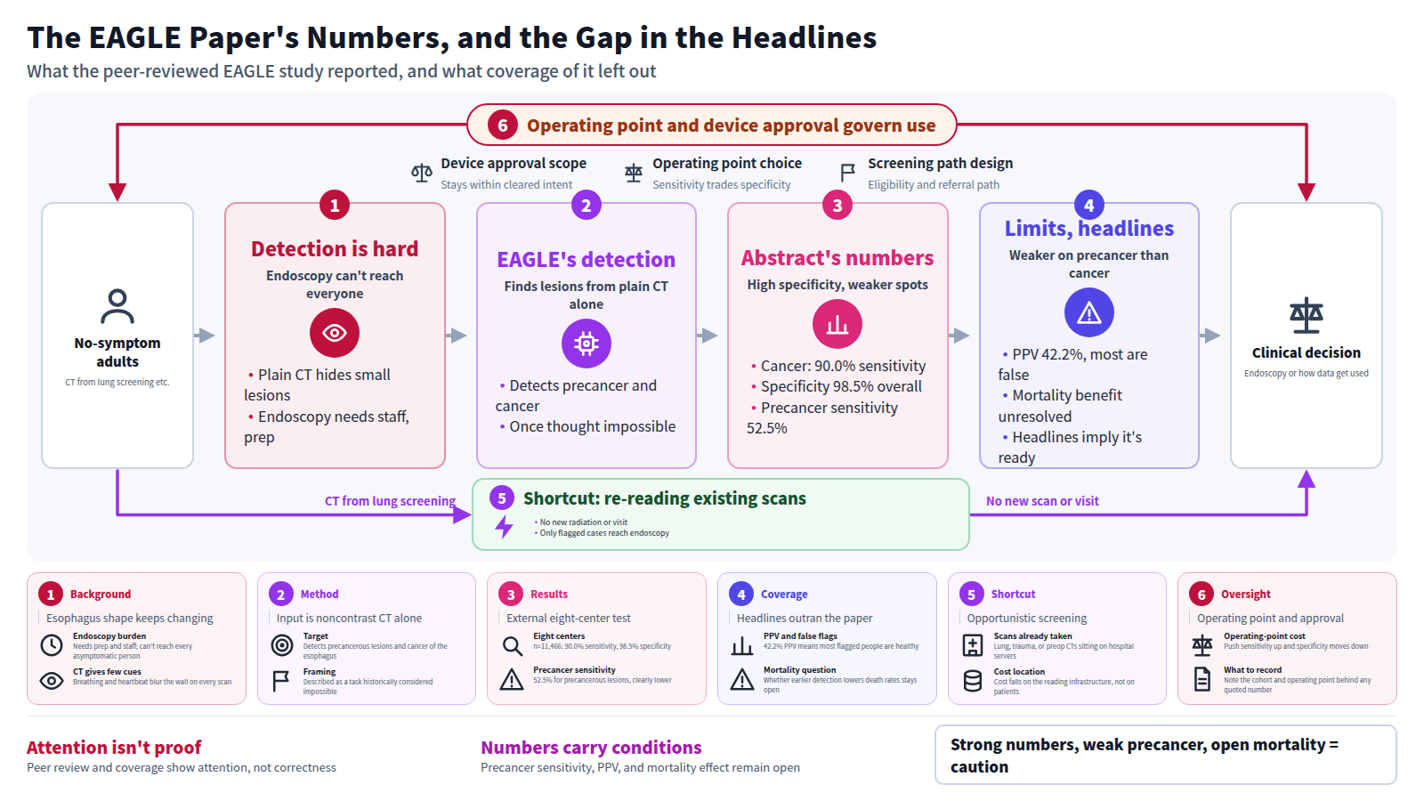
Why early esophageal cancer is still hard to catch
The esophagus is a hollow tube. It moves with breathing and heartbeat, it collapses, and its shape changes from scan to scan. On a noncontrast chest CT, that leaves few reliable cues for telling a small early lesion from normal wall thickness. Endoscopy finds early disease reliably, but it requires preparation and staff, and it cannot be offered to every asymptomatic person.
The authors frame the problem as the absence of an accurate, noninvasive, scalable screening tool. The flip side is that noncontrast CT already sits in hospitals everywhere and is acquired in volume every day for lung screening and other indications. Images are being taken but not read for this purpose. That is where the work starts.
What the paper proposes
The authors built a model called EAGLE, for Esophageal AI-Guided malignant Lesion Evaluation. The core claim is narrow and specific: detect precancerous lesions and cancer of the esophagus from chest noncontrast CT alone. The paper describes this task as one historically considered impossible.
This work was peer reviewed and published in Nature Medicine. It is not a preprint. A strong journal and a strong reported performance are still different things from performance holding up in routine practice, and that gap is taken up below.
What was shown, within the abstract
Training used data from two centers and 6,813 patients. Validation spanned 12 centers in three countries and 80,612 patients, covering both opportunistic reading of existing scans and population-based screening.
- Multicenter external testing (eight centers, n = 11,466) reached 98.5% specificity, with 90.0% sensitivity for cancer and 52.5% for precancerous lesions.
- Low-dose CT validation (two centers, n = 1,607) gave comparable performance, which the authors read as support for screening through existing lung-cancer programs.
- Calibration in a real-world cohort (three centers, n = 35,402) cut false positives by 72.7% while preserving sensitivity.
- Prospective hospital validation (n = 17,446) reached a 42.2% positive predictive value; real-world low-dose screening (n = 10,959) reached 99.94% specificity.
- In paired CT-endoscopy cohorts (two centers, n = 702), a higher-sensitivity operating point gave 65.0% for precancerous lesions and 78.4% for stage I disease.
These are reported results within the scope of this paper, and claims made by its authors.
A concrete look: two ways to deploy it
The same model means different things depending on where it is placed. The abstract distinguishes two settings.
Re-reading scans already acquired
Chest noncontrast CTs taken for other reasons — lung assessment, trauma, preoperative workup — accumulate on hospital servers. Pointing the model at them adds no new radiation and no new visit. Only flagged individuals go to endoscopy. The cost falls on the reading infrastructure.
Scanning people for screening
Low-dose CT is acquired in a screening program and read there. Eligibility rules, acquisition parameters, and the referral path to endoscopy all have to be designed first. New radiation and new cost are incurred, so the burden of demonstrating benefit is heavier.
The abstract separates opportunistic and population-based settings precisely because this difference changes how the numbers should be read.
What is not new, and where the limits are
Deep learning models that detect lesions in images are not new. What is new here is the combination of target and input — reading the esophagus off noncontrast CT — rather than the methodological frame itself.
The limits are already visible in the reported numbers. Sensitivity for precancerous lesions is clearly lower than for cancer. If catching disease at the precancerous stage is the main value of screening, that is exactly where the model is weakest. The positive predictive value also means that more than half of flagged individuals turn out to have nothing on endoscopy. In a screening setting, those false positives consume both patient anxiety and endoscopy capacity.
Much else cannot be read from the abstract at all. The distribution of scanner models and acquisition parameters across validating centers, the age and baseline risk of the populations, how completely endoscopic confirmation was performed, and the length of follow-up are not stated there. Esophageal cancer incidence varies sharply by region, so how the positive predictive value would move in a population with different prevalence is likewise outside what the abstract shows. The largest open question is whether earlier detection reduces mortality; the authors themselves keep this to an exploratory suggestion that referring high-risk individuals for endoscopy could improve screening efficiency.
What the coverage said, and where it diverges
Beyond the journal's own news page, several independent outlets picked the work up. Lining up the headlines shows how the message shifted.
- The journal-side headline stays close to the paper's title, describing large-scale esophageal cancer screening through noncontrast CT and artificial intelligence[2].
- One general outlet framed it as frontier AI leaving the lab to change early cancer screening globally[3].
- Another covered a related current as an early screening method for pancreatic cancer[4], with the target organ swapped out entirely.
The divergence is clear. The paper states that detection performance reached a given level. It does not state that the method is ready to serve as screening worldwide. Sensitivity for precancerous lesions, positive predictive value, and the unresolved mortality question all drop out of the headlines. Passing peer review and being picked up by several independent outlets shows the work drew attention; neither fact is by itself evidence of correctness or of clinical value.
What this connects to in pharma and regulatory work
Three connections stand out. First, the mix of patients being detected changes. If a larger share is caught as precancerous lesions or stage I disease, the stage distribution of treatable patients shifts, and with it the assumptions behind any therapy aimed at early disease or the perioperative setting.
Second, trial enrollment. A cohort surfaced by an imaging model differs in background from a cohort that presented with symptoms. When "flagged by an AI reading" becomes part of the enrollment path, generalizability cannot be explained without stating which device and which operating point produced the flag.
Third, regulatory status. Software that supports diagnosis is regulated as a medical device. Performing well and being used within an authorized intended use are separate matters, and moving the operating point trades sensitivity against specificity. The abstract explicitly reports a higher-sensitivity operating point, which makes the choice of setting a clinical decision that has to be documented and recorded. Anyone quoting these numbers in promotional or internal material has to carry the cohort and the operating point along with them.