AI and Cancer Research ── Cancer biology ── 2026-10-09
Cancer molecular biology and AI
- Abstract
Purpose: Parenchyma-sparing hepatectomy planning depends on accurate resection zones that preserve functional liver tissue without compromising oncological margins. This work investigates how different levels of anatomical and functional information complexity influence resection zone prediction for parenchyma-sparing surgical planning for primary liver cancer.
Methods: We compare three modeling paradigms: a geometric distance-based approach, an explicit perfusion-based method using vascular anatomy, and a deep learning-based model built on the U-Net architecture. All methods operate on segmentation-derived representations of the liver, tumor, and vessels. Performance is evaluated using overlap- and distance-based metrics.
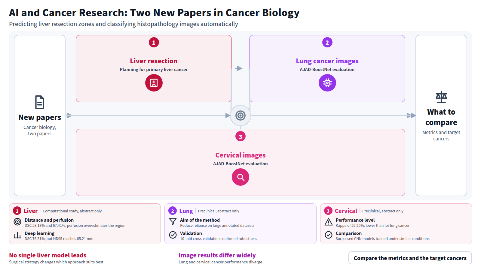
Results: The distance-based model produces predictions with limited surface deviation (HD $$_{95}$$ 95 33.89 mm) but lower overlap due to undersegmentation (DSC 58.18 %). The perfusion-based method achieves a favorable balance between overlap (DSC 67.41 %) and boundary accuracy (HD $$_{95}$$ 95 37.92 mm) but tends to overestimate the predicted region due to strict binary perfusion assumptions. The deep learning model attains the highest overlap accuracy (DSC 76.31 %) while exhibiting larger distance errors (HD $$_{95}$$ 95 65.21 mm), reflecting localized boundary inaccuracies.
Conclusion: None of the models is universally outperforming the other two for parenchyma-sparing resection planning. Deep learning shows strong predictive performance, particularly for larger resection volumes, while geometric and perfusion-based models offer greater interpretability and clinical controllability. The distance-based approach is well suited for maximal parenchyma-sparing resections, whereas perfusion-based modeling is advantageous for tumors near vessels with potential perfusion loss. Our results highlight variations due to different surgical strategies and can thus provide valuable guidance for individual patient’s surgery planning.
Journal IF-equivalent: 3.0 (OpenAlex 2-year mean citedness, value as of 2026-10-08, retrieved 2026-10-09; not the official Clarivate IF)Reference: Rothert J, Rakshit J, Salz JL, Huettl F, Ehses V, Huber T, et al. Resection zone prediction for parenchyma-sparing hepatectomy planning: a comparative study of three modeling paradigms. Int J CARS. 2026 Oct 7 [Epub ahead of print]. doi:10.1007/s11548-026-03798-7.Checked: Abstract only - Abstract
Cervical and lung cancers remain among the leading causes of cancer-related mortality worldwide.This highlights the need for accurate, scalable, and computationally efficient computer-aided diagnostic systems.Deep learning has shown remarkable success in histopathological image analysis.However, most existing approaches depend on large annotated datasets, extensive computational resources, and end-to-end supervised training.To address these limitations, this study proposes AJAD-BoostNet (Adaptive Joint Analysis and Deep Boosting Network).This novel hybrid framework integrates deep transfer learning, unsupervised clustering, dimensionality reduction, and boosting-based refinement for automated cancer histopathological image classification.The proposed framework uses a pretrained ResNet50 model to extract high-level semantic representations from histopathological images.Then, Principal Component Analysis (PCA) reduces feature dimensionality while preserving discriminative information.The optimized feature space is clustered with the K-Means algorithm for initial unsupervised classification.To further enhance predictive performance and correct clustering errors, an XGBoost-based boosting module is applied as a misclassification refinement strategy.The framework was evaluated on two benchmark datasets: the IARC Cervical Cancer Image Bank and the LC25000-based Lung Cancer Histopathological Images dataset.Experimental results demonstrate the effectiveness of the proposed approach across both datasets.For lung cancer classification, AJAD-BoostNet achieved an accuracy of 97.08%, an F1-score of 97.00%, a Cohen's Kappa score of 94.17%, and an AUC of 97.08%.For cervical cancer classification, the framework attained an accuracy of 64.84%, an F1-score of 65.00%, a Cohen's Kappa score of 29.20%, and an AUC of 64.55%.These results surpass those of conventional CNN-based models trained under similar conditions.Additionally, 10-fold cross-validation and confidence interval analyses confirmed the robustness, stability, and generalization capability of the proposed framework.The findings indicate that combining deep semantic feature extraction with clustering and boosting provides an efficient and interpretable alternative to fully supervised deep learning architectures.This makes AJAD-BoostNet a promising solution for intelligent cancer diagnosis in resource-constrained clinical environments.
Journal IF-equivalent: 0.0 (OpenAlex 2-year mean citedness, value as of 2026-10-07, retrieved 2026-10-08; not the official Clarivate IF)Reference: [No authors listed]. AJAD-BoostNet: A Hybrid Deep K-Means and XGBoost Framework for Automated Cancer Histopathological Image Classification. IJDDT. 2026 Oct 7;16(57s). doi:10.25258/ijddt.16.57s.160.Checked: Abstract only