AI and Cancer Research ── Prostate ── 2026-10-07
Prostate cancer: treatment, trials and AI
- Abstract
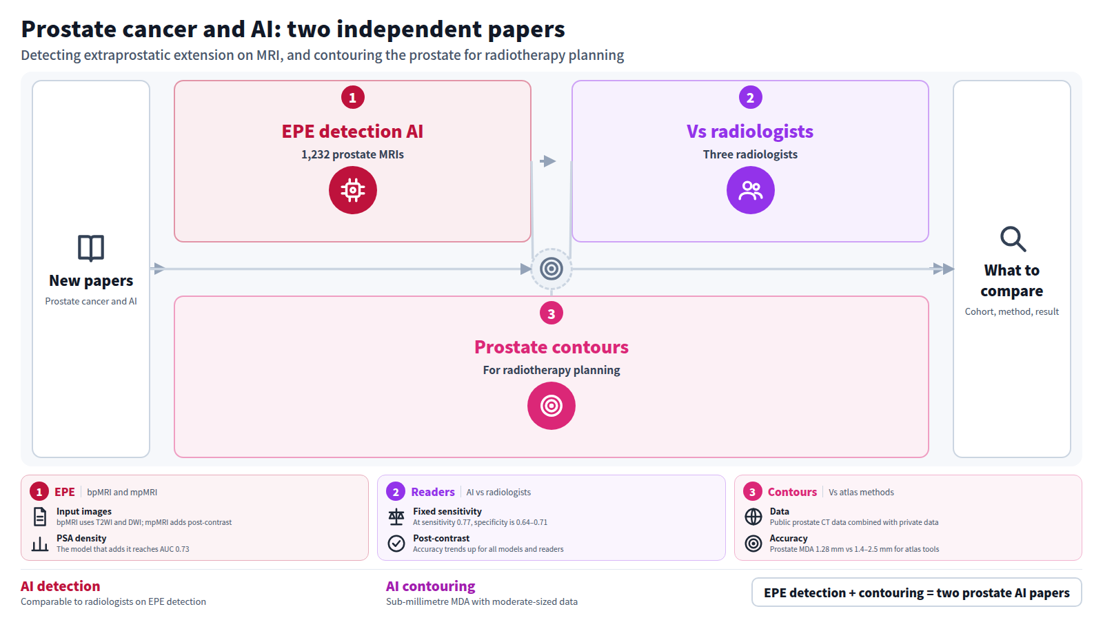
Objectives: To develop and test deep-learning models which detect extraprostatic extension (EPE) on biparametric (bpMRI) and multiparametric MRI (mpMRI) of the prostate in comparison to radiologists.
Methods: Consecutive patients at a large healthcare enterprise who underwent prostate MRI (2015 to 2023) with subsequent radical prostatectomy within 1 year were included. The dataset was divided into training/validation/test sets. Transfer learning models, composed of two multi-branch 3D convolutional neural networks based on a 3D ResNet-50 backbone, were trained on bpMRI (AI bp ) with an input of axial T2-weighted imaging (T2WI) and diffusion-weighted imaging (DWI) and on mpMRI (AI mp ) with an input of axial T2WI, axial DWI, and axial post-contrast imaging. No prostate gland or tumor segmentations were performed. A logistic regression model with prostate specific antigen density was also trained with the AI mp model (AI mpPSAD ). Three fellowship-trained abdominal radiologists evaluated the test set for EPE on a 1 to 5 scoring system based on capsular appearance, on bpMRI and mpMRI for each study. Areas under the receiver operating characteristic curves (AUC) were obtained and compared with Delong test. Diagnostic statistics were also obtained.
Results: 1,232 prostate MRIs were included (1003/113/116 in the training/validation/test sets). AUC for models were as follows: AI bp (0.71), AI mp (0.72), and AI mpPSAD (0.73). While holding sensitivity constant at the Youden index for AI bp (0.77), specificity and accuracy of the AI bp , AI mp , and AI mpPSAD were 0.64, 0.69, 0.71 and 0.71, 0.73, 0.74, respectively. The ranges of AUC for readers were: bpMRI (0.66–0.71) and mpMRI (0.69–0.74). The ranges of accuracy of the readers on bpMRI was 0.60–0.68 and on mpMRI was 0.65–0.72.
Conclusion: A segmentation-free transfer learning model performs comparably to fellowship-trained radiologists on the detection of EPE on both bpMRI and mpMRI. There was a trend of increasing accuracy with the addition of post contrast sequences for all models and readers.
Journal IF: unknown (could not be matched)Reference: Tong A, Daniels A, Ginocchio L, Smereka P, Dutt T, Umapathy L, et al. Detection of extraprostatic extension of prostate cancer by transfer learning AI models versus radiologists on biparametric and multiparametric MRI. Research Square. 2026 Oct 5 [Epub ahead of print]. doi:10.21203/rs.3.rs-11001056/v1.Checked: Abstract only - Abstract
Accurate segmentation of the prostate and surrounding structures is crucial in radiotherapy planning to ensure effective treatment delivery while minimising radiation exposure to nearby tissues. However, existing deep learning (DL) segmentation studies often exclude clinically important structures such as the penile bulb (PB) and seminal vesicles (SV), rely heavily on large proprietary datasets that are difficult to acquire in clinical settings, and lack direct comparisons with traditional atlas-based methods. To address these gaps, this study provides practical insights for hospitals considering the implementation of DL systems by evaluating 2D U-Net models across six structured experiments, including one experiment comparing a 3D U-Net model. Notably, this is the first study to utilises publicly available prostate CT data in DL training and integrates public and private datasets to improve model robustness for prostate and adjacent anatomical structures. The inclusion of underrepresented structures, such as PB and SV, enhances the clinical applicability of the models. Our results demonstrated that moderate-sized datasets, ranging from 60 to approximately 300 in our study, can achieve sub-millimetre Mean Distance Agreement (MDA) for key organs at risk like the bladder (0.60 mm) and rectum (0.93 mm), reducing reliance on large-scale annotated data. Models trained on routine clinical contours performed well without extensive manual refinement, demonstrating that existing clinical data can be leveraged for model training. In comparisons with four commercial atlas-based tools, the 2D U-Net models achieved superior accuracy for key anatomical structures, including the prostate, bladder, and SV, with our deep learning model achieving an MDA of 1.28 mm for the prostate compared to the 1.4-2.5 mm range of the atlas tools. Although 3D models provided improved spatial context for small structures such as SV, 2D models proved to be a practical alternative due to lower computational demands, making them suitable for resource-limited clinical settings.
Journal IF-equivalent: 2.4 (OpenAlex 2-year mean citedness, value as of 2026-10-06, retrieved 2026-10-07; not the official Clarivate IF)Reference: Hizam DA, Saad M, Ung NM, Muaadz A, Salleh FM, Tan LK. Practical deep learning solutions for prostate cancer segmentation and implementation in radiotherapy planning. Phys Eng Sci Med. 2026 Oct 5 [Epub ahead of print]. doi:10.1007/s13246-026-01815-2.Checked: Abstract only